
Square Root Sign in Constrictive Pericarditis
The square root sign in constrictive pericarditis is a characteristic dip-and-plateau pattern on ventricular pressure tracings during cardiac catheterization. It occurs when rapid early diastolic filling is followed by an abrupt limitation of ventricular expansion caused by a stiff, noncompliant pericardium.
This finding helps clinicians recognize constrictive physiology, but it is not diagnostic on its own. Understanding the relationship between ventricular pressure, diastolic filling, and respiratory changes is essential for distinguishing constrictive pericarditis from restrictive cardiomyopathy.
Medical review: This article requires review by a qualified cardiologist before publication.
Last updated: September 29, 2026.
Key Takeaways
- The square root sign is also called the dip-and-plateau pattern.
- It is identified on ventricular pressure tracings during cardiac catheterization.
- The pattern reflects rapid early diastolic filling followed by restricted ventricular expansion.
- Right and left ventricular end-diastolic pressures may be elevated and nearly equal.
- Respiratory ventricular pressure discordance is an important finding that supports constrictive pericarditis.
- The square root sign can also occur in restrictive cardiomyopathy, so it cannot confirm the diagnosis by itself.
What Is the Square Root Sign in Constrictive Pericarditis?
The square root sign is a characteristic change in ventricular pressure during diastole. It begins with a rapid pressure decline and early ventricular filling, followed by a sudden rise and a relatively flat plateau. The resulting waveform resembles the mathematical square root symbol.
In constrictive pericarditis, the pericardium becomes stiff and restricts the heart’s ability to expand. Blood initially enters the ventricles rapidly, but further filling becomes limited as the heart reaches the volume allowed by the rigid pericardium. This produces the characteristic dip-and-plateau pressure pattern.
Why Is It Called the Dip-and-Plateau Pattern?
The name describes the two main components of the ventricular pressure waveform. The initial dip represents early diastolic relaxation and rapid filling. The subsequent plateau reflects the abrupt limitation of further ventricular expansion.
The pattern is particularly useful during invasive hemodynamic evaluation. However, similar waveforms can occur in other conditions associated with impaired ventricular filling, including restrictive cardiomyopathy.
Hemodynamic Findings in Constrictive Pericarditis
Constrictive pericarditis produces characteristic changes in cardiac filling pressures. The rigid pericardium limits total cardiac volume, causing elevated diastolic pressures and exaggerated interactions between the ventricles.
Cardiac catheterization can reveal several findings, including the square root sign, elevated right ventricular end-diastolic pressure, and near-equalization of right and left ventricular filling pressures. Clinicians interpret these measurements alongside respiratory pressure changes and imaging findings.
Diastolic Pressure Equalization
In constrictive pericarditis, right and left ventricular end-diastolic pressures are often elevated and may differ by approximately 5 mmHg or less.
This occurs because the stiff pericardium restricts expansion of both ventricles within a relatively fixed volume. However, pressure equalization is not specific to constrictive pericarditis and may also occur in other cardiac conditions.
Commonly Cited Hemodynamic Criteria
The following measurements are commonly discussed in the differential diagnosis of constrictive pericarditis and restrictive cardiomyopathy.
| Hemodynamic finding | Commonly cited reference |
| Right and left ventricular end-diastolic pressure difference | Approximately 5 mmHg or less |
| Right ventricular end-diastolic pressure | Often at least one-third of RV systolic pressure |
| Right ventricular systolic pressure | Usually below approximately 50 mmHg in classic constriction |
| Systolic area index | Greater than 1.1 supports constrictive physiology |
| Ventricular pressure waveform | Dip-and-plateau pattern may be present |
These are traditional clinical reference criteria, not universal diagnostic cutoffs. Individual measurements vary with the patient’s condition, volume status, and measurement technique. The complete clinical and hemodynamic picture is more important than any single threshold.
How Is the Square Root Sign Identified During Cardiac Catheterization?
Cardiac catheterization allows clinicians to measure pressure directly inside the heart. During the procedure, pressure tracings can be recorded from the right and left ventricles to assess ventricular filling and identify characteristic hemodynamic patterns.
The square root sign appears as an early diastolic dip followed by a rapid rise and plateau. Simultaneous pressure recordings can also help clinicians evaluate respiratory changes in ventricular systolic pressure, which are particularly useful when distinguishing constrictive pericarditis from restrictive cardiomyopathy.
What Does the Pressure Waveform Show?
The waveform begins when ventricular pressure falls during early diastole. Blood enters the ventricle rapidly, producing an initial decline in pressure followed by a sharp increase as further filling becomes restricted.
The later plateau represents the limited capacity of the ventricles to accommodate additional blood. This pattern reflects the mechanical effects of pericardial constriction rather than a specific abnormality in the mathematical square root symbol itself.
Respiratory Hemodynamics and Ventricular Interdependence
Respiratory hemodynamics are particularly important in diagnosing constrictive pericarditis. The rigid pericardium restricts the heart’s total volume, making the two ventricles more dependent on one another during changes in venous return.
During inspiration, right ventricular filling increases while left ventricular filling typically decreases. This produces opposite respiratory changes in right and left ventricular systolic pressure. The phenomenon is called ventricular pressure discordance and supports the diagnosis of constrictive physiology.
Systolic Area Index
The systolic area index is a hemodynamic measurement used to assess enhanced ventricular interdependence. It compares the ratio of right ventricular to left ventricular systolic pressure-time areas during inspiration with the corresponding ratio during expiration.
A systolic area index greater than 1.1 is a commonly cited threshold supporting constrictive pericarditis. It reflects the exaggerated interaction between the ventricles during respiration.
This measurement requires appropriate simultaneous ventricular pressure recordings and should be interpreted by clinicians familiar with invasive hemodynamic testing.
Ventricular Pressure Discordance
Ventricular pressure discordance refers specifically to opposite respiratory changes in right and left ventricular systolic pressures.
During inspiration, right ventricular systolic pressure generally increases while left ventricular systolic pressure decreases. During expiration, the relationship reverses. This pattern results from exaggerated ventricular interdependence within the restricted pericardial space.
It is an important distinction from restrictive cardiomyopathy, where ventricular systolic pressure changes are typically concordant rather than discordant.
Clinical Signs and Symptoms of Constrictive Pericarditis
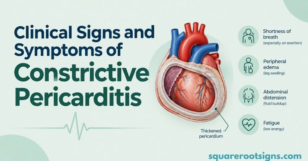
Constrictive pericarditis commonly presents with symptoms of systemic venous congestion and impaired cardiac filling. Patients may experience fatigue, breathlessness, abdominal swelling, and swelling of the legs.
Physical examination can reveal elevated jugular venous pressure, a prominent y descent, a positive Kussmaul sign, and a pericardial knock. These findings can support clinical suspicion, but none is sufficient by itself to establish the diagnosis.
Kussmaul Sign
Kussmaul sign is a paradoxical increase or failure of the jugular venous pressure to decrease during inspiration. It occurs when the right side of the heart cannot accommodate the normal increase in venous return.
Although Kussmaul sign is associated with constrictive pericarditis, it can also occur in restrictive cardiomyopathy and other conditions that impair right ventricular filling.
Pericardial Knock
A pericardial knock is an early diastolic heart sound associated with abrupt cessation of ventricular filling. It may be heard in patients with constrictive pericarditis and can resemble other extra heart sounds.
The sound results from sudden restriction of ventricular expansion. It is a useful clinical clue when considered alongside symptoms, physical examination, and diagnostic imaging.
Prominent Jugular Venous y Descent
The jugular venous y descent reflects rapid emptying of the right atrium into the right ventricle during early diastole.
In constrictive pericarditis, early ventricular filling may initially be rapid. The subsequent restriction of ventricular expansion produces characteristic changes in cardiac filling and venous pressure waveforms. A prominent y descent can therefore support the diagnosis.
Imaging Findings in Constrictive Pericarditis
Imaging plays an essential role in identifying constrictive physiology and assessing the pericardium. Echocardiography is generally the initial imaging test, while cardiac magnetic resonance and computed tomography can provide additional information.
Importantly, a normal pericardial thickness does not exclude constrictive pericarditis. Diagnosis requires consideration of clinical symptoms, hemodynamic findings, and imaging evidence.
Echocardiography
Echocardiography can reveal respiratory changes in ventricular filling, abnormal interventricular septal motion, and changes in mitral and tricuspid inflow velocities.
Other findings include increased medial mitral annular e′ velocity, annulus reversus, a dilated inferior vena cava, and expiratory hepatic vein diastolic flow reversal. A combination of these findings can support the diagnosis.
Cardiac MRI and CT
Cardiac MRI can help assess pericardial thickening, inflammation, ventricular interaction, and myocardial abnormalities. It is particularly useful when clinicians need to distinguish constrictive pericarditis from restrictive myocardial disease.
Cardiac CT is especially useful for detecting pericardial calcification and assessing pericardial anatomy. However, calcification alone does not prove that constriction is present.
Constrictive Pericarditis vs. Restrictive Cardiomyopathy
Constrictive pericarditis and restrictive cardiomyopathy can produce similar symptoms and elevated ventricular filling pressures. Both may also demonstrate a square root sign during cardiac catheterization.
The principal difference is the location of the abnormality. Constrictive pericarditis results from a restrictive pericardium, whereas restrictive cardiomyopathy results from abnormal stiffness of the heart muscle.
| Feature | Constrictive pericarditis | Restrictive cardiomyopathy |
| Primary abnormality | Stiff or restrictive pericardium | Stiff myocardium |
| Square root sign | May be present | May also be present |
| Diastolic pressure elevation | Common | Common |
| Diastolic pressure equalization | Often present | May occur |
| Respiratory ventricular pressure changes | Typically discordant | Typically concordant |
| Ventricular interdependence | Exaggerated | Usually less pronounced |
| Pericardial thickening | May be present | Not a defining feature |
| Pericardial calcification | May be present | Not characteristic |
| Mitral annular e′ velocity | Often preserved or increased medially | Often reduced |
| Main treatment approach | Depends on cause and reversibility; may require pericardiectomy | Treat underlying myocardial disease and associated heart failure |
Respiratory ventricular pressure discordance and imaging evidence of pericardial constriction are particularly useful in differentiating these conditions. However, mixed disease can occur, and specialist evaluation may be necessary.
Causes of Constrictive Pericarditis
Constrictive pericarditis can develop after several types of pericardial injury or inflammation. The most common causes vary according to geography, clinical setting, and the patient’s medical history.
In the United States and other developed countries, idiopathic or presumed viral disease, previous cardiac surgery, and chest radiation are important causes. Tuberculosis remains a major cause in many regions where the infection is endemic.
Other causes include bacterial pericarditis, connective tissue disorders, malignancy, trauma, and certain inflammatory conditions.
Treatment and Clinical Management
Treatment depends on the cause, duration, severity, and reversibility of the pericardial disease.
Some patients have transient constrictive pericarditis associated with active inflammation. These patients may improve with appropriate anti-inflammatory treatment and careful clinical monitoring.
Patients with persistent chronic constriction caused by a rigid, fibrotic pericardium may require surgical pericardiectomy. Diuretics can help relieve fluid retention and congestion in selected patients, but they do not remove the underlying mechanical restriction.
Treatment decisions require specialist assessment because the risks and benefits of surgery depend on the patient’s overall condition and the underlying cause.
Conclusion
The square root sign in constrictive pericarditis is a characteristic dip-and-plateau pattern that reflects rapid early ventricular filling followed by abrupt restriction. It is an important finding during cardiac catheterization, but it cannot independently establish the diagnosis.
Elevated and nearly equal ventricular end-diastolic pressures, respiratory ventricular pressure discordance, and supportive echocardiographic or cardiac imaging findings help clinicians distinguish constrictive pericarditis from restrictive cardiomyopathy. Accurate diagnosis requires interpreting these findings together with the patient’s clinical presentation.
FAQs
1. What are the radiological signs of constrictive pericarditis?
The main radiological signs are pericardial calcification on chest X-ray or CT and pericardial thickening on CT or MRI. Echocardiography may show septal bounce and a dilated inferior vena cava.
2. What is the hallmark sign of pericarditis?
The hallmark ECG finding of acute pericarditis is widespread, concave ST-segment elevation with PR-segment depression.
3. What is a positive Kussmaul sign?
A positive Kussmaul sign is a rise or failure of the jugular venous pressure to fall during inspiration. It is commonly associated with constrictive pericarditis.
4. Can Friedreich’s sign be a sign of constrictive pericarditis?
Yes. Friedreich’s sign is a rapid, prominent descent in the jugular venous pulse. It can be seen in constrictive pericarditis due to rapid early ventricular filling.

Jack Harrison is a contributor at Square Root Signs, sharing creative ideas, useful information, and engaging content to help readers discover something valuable with every visit.





One Comment